Progress evolves on rapid testing


Historically, methodology to detect pathogens in the food supply that have the potential to make people sick took four to five days. When processors are dealing with perishable products, long, time-consuming testing is not really helpful to the industry. This is why rapid testing is used to screen products for pathogens in a timely manner and in less than 24 hours, which most rapid methods are.
The heaviest area for rapid testing is used to detect O157:H7 Shiga toxin-producing E. coli (STEC) pathogens during the screening of raw materials used to make ground beef, says Mohammad Koohmaraie, chief executive officer of the meat division of IEH Laboratories & Consulting Group, in Lake Forest Park, Wash. Combos of trimmings are mandated to be lotted (usually 2,000 pounds but not more than 10,000 pounds), sampled and tested for E. coli O157. The sample must be negative before being released into commerce. Because testing beef trim occurs at the end of the process, some beef processors show interest in rapid testing methods being used earlier in the process to assess the entire process, Koohmaraie says.
The most recent developments to rapid testing are occurring with the six non-O157 STEC pathogens in the beef industry, which were declared adulterants by U.S. Department of Agriculture (USDA) in September 2012, Koohmaraie says. As such, the primary objective of researchers in the coordinated agricultural program (CAP) grant from the USDA is to research detection methods for the six non-O157 STEC pathogens in the beef industry.
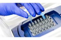
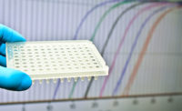
In terms of detection, processors want to quantify the number of organisms in a sample. Polymerase chain reaction (PCR), a molecular-based rapid test, can perform this feat on hundreds of samples at a time in real time, says T.G. Nagaraja, university distinguished professor in the Department of Diagnostic Medicine/Pathobiology in the College of Veterinary Medicine at Kansas State University in Manhattan and a researcher in the CAP grant. Nagaraja’s laboratory has developed three sets of PCR assays, detecting up to 10 genes, to quantify O157 plus the top six non-O157 STEC. The PCR assays can detect the O group, Shiga toxin-producing genes, and the intimin protein (encoded by the eaegene), all necessary to be present to be pathogenic for humans. The PCR test can determine whether these genes are present, and if a sample is positive, quantify the concentration per gram.
“The concentration is very important for the risk of the assessment of the STEC in cattle,” Nagaraja says. “They like to quantify the risk based on concentration, because the higher the concentration, the higher the risk.”
PCR testing also has benefits in accessing the efficacy of interventions. For example, one could take a sample before spraying a chemical intervention on a hide to determine the concentration of bacteria on a square centimeter basis. Another sample could be obtained after the intervention to determine the reduction, Nagaraja explains.
PCR does have a limitation in that it can indicate whether a sample has an O group and Shiga toxin genes present, but not whether they are together, i.e., the Shiga toxin genes could be with O157 or another bacteria. Nagaraja’s laboratory is also looking at a culture method to quantify the concentration of the organisms in a sample, for which culture methods are tedious and labor intensive. An alternative to this would be to use spiral plating, a technique used in microbiology for more than 30 years. Spiral plating would deposit the sample on a plate that spins, so one would know exactly what concentration of the sample was deposited on each area of the plate. Nagaraja’s laboratory used spiral plating to validate the quantity of the centration of the O group in a fecal sample, count the colonies and pick the colonies along with identifying whether it has a virus gene.
“Spiral plating could be used to quantify Shiga toxin-producing E. coli, not just the O group, which PCR does,” Nagaraja explains. “We tested that and we have shown that it works, so we are in the process of preparing a manuscript for submission.”
The CAP grant also continues to work with another PCR-type method called multiplex oligonucleotide ligation-PCR (MOL-PCR), which seeks to increase the number of unique targets for detection of non-O157 STEC. MOL-PCR detects specific DNA sequences in a multiplex fashion with high-throughput and the ability to work in a complex background.
Researchers in the CAP grant also continue research into monoclonal antibody development, which will have the ability to detect the part of the molecule that makes it specific for the O group, says Rod Moxley, a professor at the School of Veterinary Medicine and Biomedical Sciences at the University of Nebraska-Lincoln and project director of the CAP grant. Researchers are using a waveguide-based optical sensor that targets the proteins or complex carbohydrates of STEC in a rapid fashion.
The monoclonal antibodies developed would then be used for further test development. For example, lateral flow assays are used in conjunction with antibodies to detect the bacteria. A limitation to lateral flow is that each antibody detects one specific type of molecule, for example the O26 antigen, Shiga toxin or intimin protein. If the assay is intended specifically to detect the target organism in a sample, all three molecules would need to be detected and known to come from the same bacterial cell, Moxley explains.
Nanotechnology or microfluidics is also being experimented with to test extremely small volumes of material down to one nanoliter, with a goal to capture and isolate one bacterial cell from a given concentration of cells in suspension for testing, Moxley explains.
“This could be a key to future test development, because the problem with needing to detect all these different targets on one bacterial cell could be potentially overcome through microfluidics,” he says.
Moxley cites a recent report that used a microfluidic device to detect O157 E. coli. Monoclonal antibodies were used in this case for the capture and detection of antigens on the surfaces of bacteria. “Microfluidic work helps with rapid testing because they are dealing with extremely small volumes, which allows for reactions to take place in an accelerated format because it’s working on one tiny area,” Moxley explains.
Ultimately, these rapid tests and combinations of testing are looking for ways to try to minimize background organisms, i.e., trying to get these organisms by themselves in one place for their detection, Moxley says.
With the quest to find something better, Koohmaraie cautions that the tests may lose accuracy.
“I was a proponent years ago of this approach as well: That is, let’s look to find out what makes an E. coli pathogenic,” he explains. “What are the genes that make bacteria pathogenic, and start a screen for these genetic targets. I have changed my opinion, because I believe the more precise we get, the more the possibility that we may lose the ability to detect some other E. coli strains that could be pathogenic because they may not have the particular genetic targets. To me, the wider the net is cast the better chance of catching pathogenic E. coli and eliminating the possibility of releasing contaminated products into commerce. The only drawback to this approach is the possibility that products may be designated as positive that are not. My main concern is false negatives, as the consequences of releasing positive products could have devastating consequences to the consuming public and the producing company alike.” NP
Looking for a reprint of this article?
From high-res PDFs to custom plaques, order your copy today!


